Soubor:Reconstructed Spanish Flu Virus.jpg
Původní soubor (2 126 × 1 312 pixelů, velikost souboru: 664 KB, MIME typ: image/jpeg)
| Tento soubor pochází z Wikimedia Commons. Níže jsou zobrazeny informace, které obsahuje jeho tamější stránka s popisem souboru. |
Popis
| Popis |
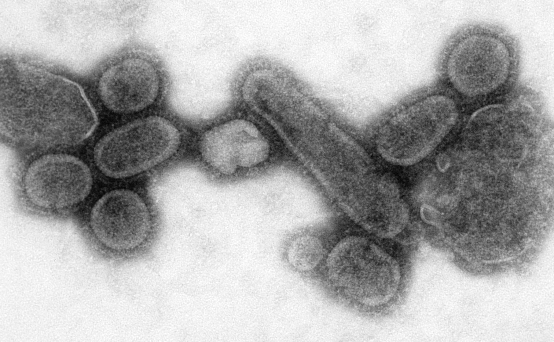
English: This negative stained transmission electron micrograph (TEM) showed recreated 1918 influenza virions that were collected from the supernatant of a 1918-infected Madin-Darby Canine Kidney (MDCK) cell culture 18 hours after infection.
In order to sequester these virions, the MDCK cells were spun down (centrifugation), and the 1918 virus present in the fluid was immediately fixed for negative staining. Dr. Terrence Tumpey, one of the organization’s staff microbiologists and a member of the National Center for Infectious Diseases (NCID), recreated the 1918 influenza virus in order to identify the characteristics that made this organism such a deadly pathogen. Research efforts such as this, enables researchers to develop new vaccines and treatments for future pandemic influenza viruses. The 1918 Spanish flu epidemic was caused by an influenza A (H1N1) virus, killing more than 500,000 people in the United States, and up to 50 million worldwide. The possible source was a newly emerged virus from a swine or an avian host of a mutated H1N1 virus. Many people died within the first few days after infection, and others died of complications later. Nearly half of those who died were young, healthy adults. Influenza A (H1N1) viruses still circulate today after being introduced again into the human population in the 1970s.Deutsch: Virionen des rekonstruierten Virus der spanischen Grippe, 18 Stunden nach Infektion der Kultur.
Français : Virus reconstitué de la grippe espagnole, celui qui est le plus proche par ses effets sur l'organisme du virus H5N1. Photographie électronique du Virus de 1918 rétrospectivement reconstitué par génie génétique à partir d'échantillons de restes humains de 1918.
日本語: 患者の遺体から見つかったゲノムより復元されたスペインかぜウイルス.
Italiano: Immagine al microscopio elettronico del virus del 1918 (ricreato in laboratorio).
Português: Vírus da gripe espanhola reconstituído.
Español: Imagen al microscopio electrónico del virus de 1918 (recreado en laboratorio).
中文:重建的西班牙流感病毒。.
Türkçe: Yeniden yaratılmış 1918 influenza virüsü.
Magyar: A spanyolnátha egykori áldozataiból nyert mintából kitenyésztett kórokozó elektronmikroszkópos képe.
עברית: מבנה נגיף השפעת הספרדית
한국어: 스페인 독감 바이러스의 사진.
Íslenska: Endurgerður vírus sem ræktaður var úr manni sem lést árið 1918.
Latina: Imago cum microscopio viri.
Sunda: Wangun ulang virus flu 1918. |
||
| Datum | |||
| Zdroj |
|
||
| Autor |
|
||
| Svolení (Užití tohoto souboru) |
PD-USGov-HHS-CDC English: None - This image is in the public domain and thus free of any copyright restrictions. As a matter of courtesy we request that the content provider be credited and notified in any public or private usage of this image. |
Licence
Tento obrázek je dílem zaměstnance Centers for Disease Control and Prevention, který je součástí United States Department of Health and Human Services, pořízeným nebo vytvořeným v rámci jeho pracovní náplně. Jako dílo federální vlády USA je tento obrázek volným dílem.
|
Historie souboru
Kliknutím na datum a čas se zobrazí tehdejší verze souboru.
| Datum a čas | Náhled | Rozměry | Uživatel | Komentář | |
|---|---|---|---|---|---|
| současná | 22. 10. 2018, 06:09 | | 2 126 × 1 312 (664 KB) | Opencooper | higher res |
| 23. 2. 2006, 04:35 |  | 700 × 431 (45 KB) | Mnh | This negative stained transmission electron micrograph (TEM) showed recreated 1918 influenza virions that were collected from the supernatant of a 1918-infected Madin-Darby Canine Kidney (MDCK) cell culture 18 hours after infection. In order to sequester |
Využití souboru
Tento soubor používá následující stránka:
Globální využití souboru
Tento soubor využívají následující wiki:
- Využití na ar.wikipedia.org
- Využití na ast.wikipedia.org
- Využití na az.wikipedia.org
- Využití na ca.wikipedia.org
- Využití na de.wikipedia.org
- Využití na de.wikinews.org
- Využití na en.wikipedia.org
- Využití na en.wikiquote.org
- Využití na es.wikipedia.org
- Využití na fr.wikipedia.org
- Využití na gl.wikipedia.org
- Využití na he.wikipedia.org
- Využití na hr.wikipedia.org
- Využití na hu.wikipedia.org
- Využití na hy.wikipedia.org
- Využití na is.wikipedia.org
- Využití na it.wikipedia.org
- Využití na ja.wikipedia.org
- Využití na ko.wikipedia.org
- Využití na la.wikipedia.org
- Využití na lfn.wikipedia.org
- Využití na lij.wikipedia.org
- Využití na lmo.wikipedia.org
- Využití na nl.wikipedia.org
- Využití na nv.wikipedia.org
- Využití na pt.wikipedia.org
- Využití na ro.wikipedia.org
Zobrazit další globální využití tohoto souboru.